
The dental industry has seen a revolution with the introduction of advanced technologies, such as cone beam computer tomography and intraoral cameras for 3D imaging. Both of these technologies play a complementary role and are readily used by an emergency dentist Burlingame.
Cone beam computed tomography with intraoral cameras plays a vital role in diagnostic capabilities, treatment planning, and ensuring oral care. With these techniques, an emergency dentist provides effective and efficient management of dental issues. It is important to understand these two concepts for dental treatments:
Cone-beam Computed Tomography (CBCT) in 3D Imaging
CBCT is a state-of-the-art imaging technology that has brought the use of three-dimensional scanning techniques in dentistry. The image of the affected teeth with gums and jaws can be achieved which can provide a more comprehensive view than any traditional methods. It allows dentists to diagnose and treat complex issues such as knocked-out teeth, fractures, root decay, etc.
CBCT is the visual aid for a patient to get treated in case of temporomandibular joint (TMJ) disorders with greater accuracy. For dental trauma like this, CBCT can reveal detailed reports of the extent of damage and henceforth guide emergency dental prosthetics in determining a customized and effective treatment schedule for the patient. Dentists can monitor progress and make real-time adjustments.
CBCT can also help a person to understand the dental condition in a detailed manner. The 3D images can be taken and restored with the help of a Computer-Aided Design technique to determine the bone structure and density. The affected area or tooth can be viewed from different angles, allowing the dental surgeon to explain the loss of soft tissues, and internal anatomy of teeth and identify issues that might not be visible in traditional X-ray images.
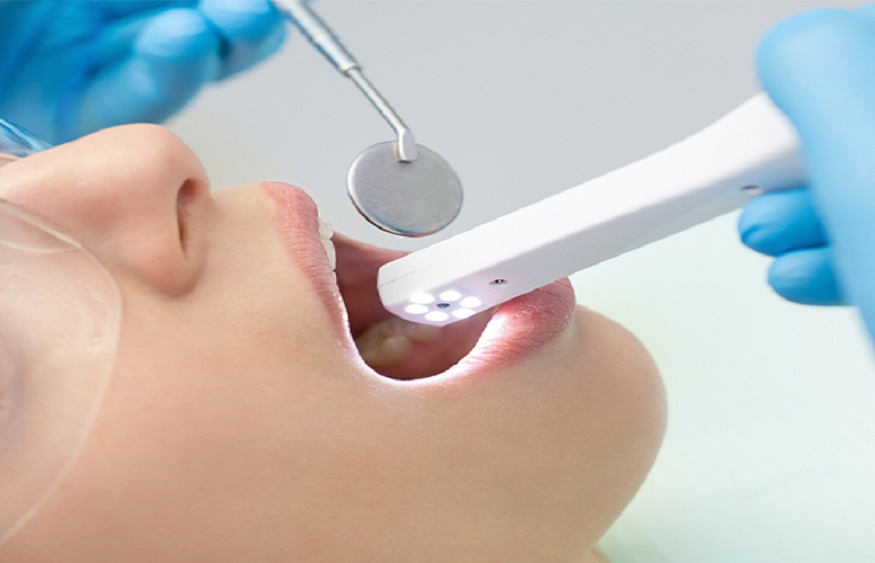
Intraoral Cameras for High-Resolution Dental Images
Few handheld devices can be adjoined with a camera to take oral images. They can capture high-resolution images that can be displayed on a monitor for getting a closer look and detailed analysis reporting can be prepared. Both the dentist and patient can look at the images and understand the procedure of dental surgery and restoration.
The hidden issues underlying the jaw, gums, or root of a tooth can be easily determined with the help of an intraoral camera. Minute cracks and infections in the deep tissues of gum can be easily recognized and explained to the patients by showing real-time images of their dental condition. Dentists can explain the issues and suggest effective treatment plans accordingly.
The images and reports can be documented by the doctor and the patient for adhering to the immediate care and follow-up treatments. The progress in the dental condition can also be monitored regularly by the usage of intraoral cameras. The images can be saved and used for comparison for adjusting to the treatment plan.
Importance of Advanced Technologies
Intraoral cameras are used for initial assessment followed by CBCT scans. The first one assists in identifying visible issues with ease while the second one provides an in-depth analysis of the underlying problem, revealing any hidden complications, if involved. Together they help an emergency dentist to precisely plan the dental treatment and execute them properly tracking the progression throughout.
Both of these techniques provide visual aids for dentists to maintain the 3D structure of the oral cavity and teeth. Showing patients clear and detailed images of their dental issues builds trust and helps them feel more involved in treatment decisions. CBCT and Intraoral cameras are integral parts of the modern emergency treatment provided in Burlingame ensuring the best possible care for the patients.