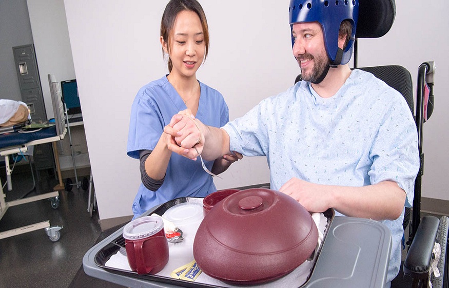
TBI is a severe and disabling condition entailing a broad range of symptoms due to a hit on the head or a sudden shake. Of all the diagnostic tools that are used in the assessment of TBI, the assessment of the pupils’ response is one of the most important. This blog covers pupillary response in Traumatic Brain Injury, the reactivity of the pupils, the pupillary light reflex, and the Neurological Pupil index (NPi).
The Relevance of Pupillary Response in TBI
The pupillary response is the condition of the pupil in terms of size and light reactions. This physiological response is an important component of neurological examinations because it offers important information about the brain’s function. Pupillary response is relevant when it comes to TBI since analyzing it helps to define the extent of the head trauma and the further treatment.
Understanding Pupil Reactivity
Pupil reactivity pertains to how the pupils respond to light and other related stimuli. Normally, the pupils of the eyes become small when there is light and become big when there is little or no light. This reaction is modulated by the ANS, the sympathetic and parasympathetic divisions of the ANS in particular.
Dysfunction of the cranial nerves increased intracranial pressure or brainstem damage can be evidenced by assessing the pupil reactivity in patients with TBI. Thus, the evaluation of the reactivity of the pupils is an important step of the neuro exam, carried out on patients with potential traumatic brain injuries.
The Pupillary Light Reflex
The pupillary light reflex is an involuntary response through which the pupils reduce in size to light. This reflex is elicited by the optic nerve also known as cranial nerve II and the oculomotor nerve also referred to as cranial nerve III. In this way, the direct and consensual reactions of the pupils are assessed based on the illumination with the light directed into the patient’s eye.
In turn, the illuminated pupil constricts. In response, the pupil of the other eye also shrinks but does not get directly illuminated by the light source. Any irregularity in these responses may point to possible neurological problems, which is why the pupillary light reflex is a useful tool in evaluating TBI.
Neurological Tools in the Assessment of TBI
Modernization in health care has brought many neurological tools that are used in the improvement of the accuracy as well as the efficiency of the TBI tests. One of the tools is the Neurological Pupil Index (NPi). Thus, the NPi can be used by the clinician as a tool to quantify the changes in the size and reactivity of the pupils.
The NPi employs infrared pupillometry; a method that captures the size and responsiveness to light of the pupils accurately. Through these measurements, the NPi puts a numerical value to the reactivity of the pupils, therefore helping to identify early neurological decline and appropriate action.
Assessment of the Neurological Status
Neurological exam is very important in the assessment of TBI patients and one of the assessments done is the pupillary response. During the exam clinicians watch the size, location, and shape of the pupils, as well as their response to light and other sources. Anything outside the norm may mean that there is more significant brain damage than is considered typical for the patient.
The neuro exam may also involve the following tests of motor and sensory function of the limbs and cranial nerves as well as reflexes and degree of cognitive impairment. These results indicate that by integrating the results of the assessment of the pupillary response with the neurological status of the patient, one can provide a more precise treatment strategy.
Conclusion
When it comes to TBI, evaluation of pupil reaction is critical to the general understanding of the condition. The degree of reactivity of the pupils and the functioning of the PLR is essential for evaluating the work of the brain and the presence of injuries. With the availability of neurological tools such as the Neurological Pupil index (NPi), the accuracy of these assessments has been enhanced and this has promoted early intervention resulting in better patient outcomes.
In the future, integration of these diagnostic methods will prove to be of significant importance as the field of research and technology progresses. Clinicians have to be careful while using these techniques while conducting an assessment for determining TBI, they have to be quite careful and precise to deliver the best of the services.

